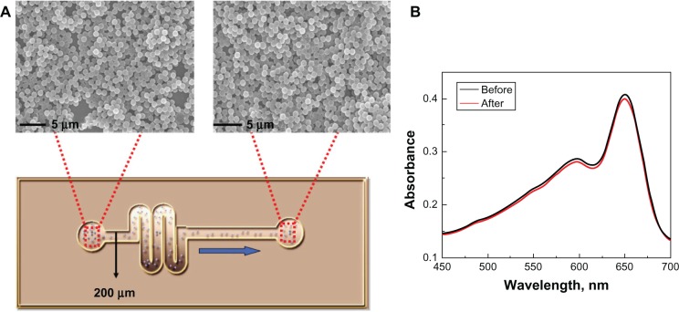
Figure 4.
(A) Scanning electron microscopy images of polystyrene polydiacetylene vesicle (PS@PDAV) microspheres before (left) and after (right) flowing through a microfluidic chip with a channel diameter of 200 μm. (B) The UV-visible absorbance spectra of PS@PDAV before (black line) and after (red line) flowing through the microfluidic chip as shown in (A).

