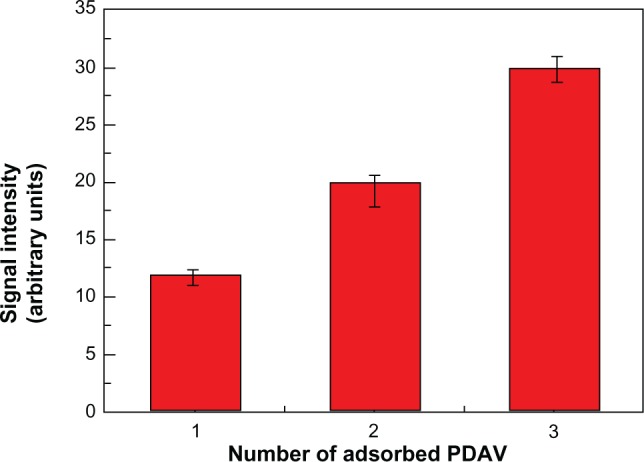
Figure 7.

Fluorescence response of polystyrene polydiacetylene vesicle (PDAV) microspheres with different PDAV quantity for H5N1 virus detection at 30 ng/mL (antibody concentration was 1:3000).
Notes: The fluorescence signal was collected by laser-scanning confocal microscopy and calculated by Image-pro plus 5.0 software (Media Cybernetics, Rockville, MD, USA). From left to right, the number of PDAV layer was one, two, and three layers.
