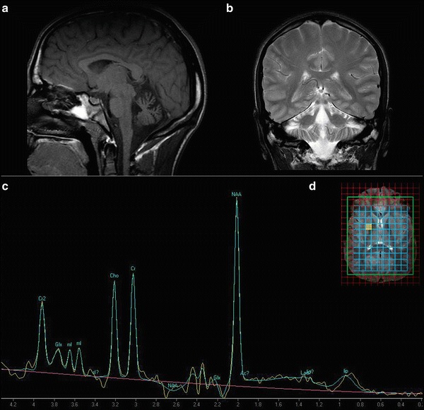
Fig. 1.

A moderate antero-superior vermian atrophy on sagittal T1-weighted (TR/TE 425 ms/10 ms) (a) and on a coronal section on T2-weighted (b) brain MRI. The scan was performed before the memantine treatment. (c) Proton MR spectroscopy (1H-MRS) at the age of 21 (clinical system of 3T, TE: 30 ms, using commercially available software) reveals no significant changes at short TE. (d) Representative location of the MRS voxels on axial T2-weighted image within the basal ganglia
