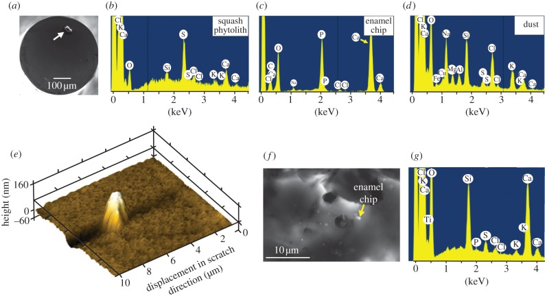
Figure 1.
(a) Scanning electron microscopy (SEM) image of a squash phytolith (arrowed) mounted on the flat-ended titanium nanoindenter tip. (b–d) Pre-test screening of chemical identity of each mounted particle by energy dispersive spectroscopy (EDS). No sputter-coating was necessary. Dust and phytoliths have distinct elemental compositions, while Ca and P peaks for enamel reflect hydroxyapatite. (e) A squash phytolith has rubbed an enamel surface, a broken fragment of which remains embedded in the enamel. Note the U-shaped trough lying to the left of the fragment. (f) Part of the surface of a quartz dust particle, post-test, littered with small enamel chips, one of which is arrowed. (g) The joint identity of quartz particle and an enamel chip was confirmed by spot EDS analysis, with the Ca and P peaks reflecting enamel, as in (c), with the other peaks mirroring those in (d).

