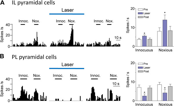
Figure 4.
Effects of optical activation on evoked activity. Effect of optical stimulation on the responses of mPFC pyramidal cells to brief (10 s) innocuous (300 g/30 mm2) and noxious (2000 g/30 mm2) somatosensory stimuli (mechanical compression of the knee, see Methods). (A) Responses of IL pyramidal cells increased during optical stimulation (1 mW, 10 Hz) in IL. Left, Peristimulus time histograms (bin width, 1 s) show responses of an individual neuron to innocuous (Innoc.) and noxious (Nox.) stimuli (horizontal bars) before, during and after laser stimulation. Right, Bar histograms show summary data (means ± SE) for the sample of IL pyramidal cells tested (n = 8). Pre, before; Post, after laser stimulation. Background activity preceding each stimulus has been subtracted from the total activity during stimulation to obtain the net evoked response. (B) Responses of PL pyramidal cells decreased during optical stimulation (5 mW, 10 Hz) in IL. Same display as in (A). Bar histograms show summary data for the sample of PL pyramidal cells tested (n = 5). * P < 0.05 (compared to control before stimulation “Pre”, Bonferroni posttests).

