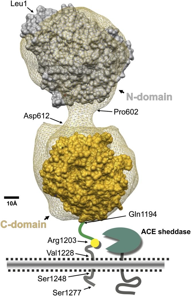
Fig. 1.
Model of human somatic ACE. This model is a three-dimensional reconstruction of the electron microscopic appearance of porcine somatic ACE (the net) combined with available human X-ray crystal structures (Chen et al., 2010; Danilov et al., 2011). It shows the ACE N-domain (Leu1–Pro601), linker region (Pro602–Asp612), C-domain (Leu613–Pro1193), stalk region (Gln1194–Arg1227), transmembrane segment (Val1228–Ser1248), and intracellular domain (Gln1249–Ser1277) (Acharya et al., 2003; Corradi et al., 2006). We also indicate the membrane-bound ACE sheddase and Arg1203, the C-terminal residue of soluble ACE.

