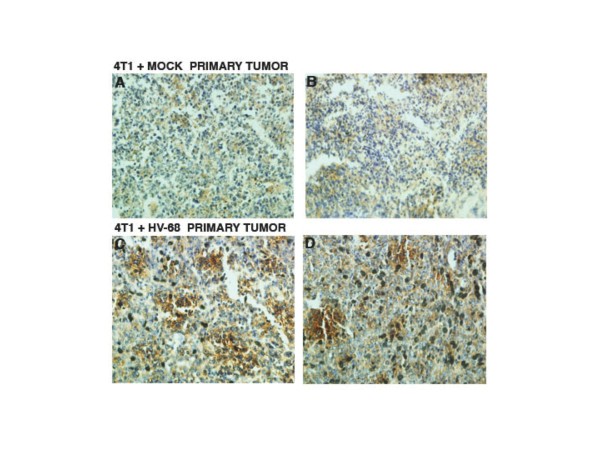
Figure 6.
VEGF-A staining of primary mammary tumor sections. At death or at day 44 following tumor cell transplantation, groups of mice (N = 10) were euthanized. Primary mammary tumors were excised from each animal, fixed in formalin, paraffin embedded, and sectioned for staining with an antibody against VEGF-A as a marker for angiogenesis. The chromogen, DAB, stains brown and was used to detect the presence of anti-VEGF-A antibody binding. Panels A and B show representative anti-VEGF-A stained microscopic mammary tumor sections from two mock treated mice transplanted with 4 T1 tumor cells. Panels C and D show representative anti-VEGF-A stained microscopic mammary tumor sections from two HV-68 infected mice transplanted with 4 T1 tumor cells.

