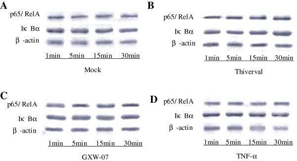
Figure 5.

Western Blot analysis of p65/RelA and IκBα in CSFV-infected PK-15 cells within 1 h. A: PK-15 cells were mock treated. B: PK-15 cells were infected with CSFV Thiverval strain at an MOI of 1. C: PK-15 cells were infected with CSFV GXW-07 strain at an MOI of 1. D: PK-15 cells were TNF-α (10ng/mL) stimulated. At 1, 5, 15, 30 min p.i, extracts of circa 20 μg total cell were prepared and subjected to Western Blot analysis with antibodies specific to p65/RelA and IκBα. Anti-β-actin served as an internal control. The experiment was repeated three times and the figure shows a representative experiment.
