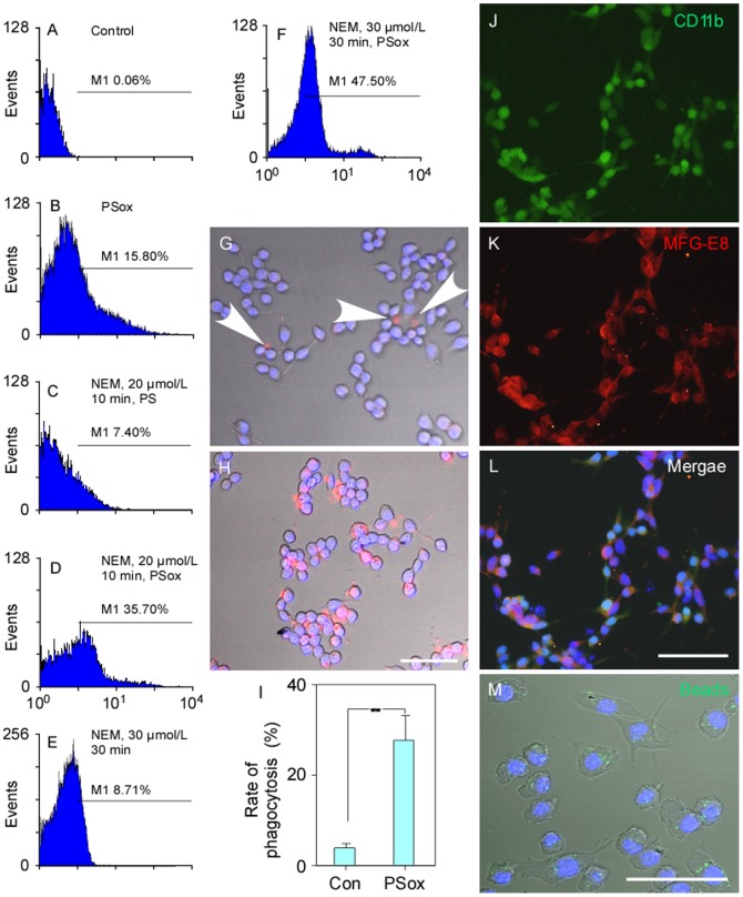
Figure 1. The detection of phosphatidylserine externalization in hRBCs labelled with Annexin V FITC fluorescence (flow cytometry) (A–F) and the phagocytosis of these cells by mouse microglial cells (G–H).
(A–F) PS externalization was (35.70%) most successful with incubation for 10 min with 20 µM NEM and then incubated with 20 µM PSox for 30 min. Although, 47.50% of hRBCs showed PS externalization after being incubated for 30 min with 30 µM NEM and then 30 min with 20 µM PSox for 30 min, cells were fragile and considered unsuitable for use. (G) Prelabled normal hRBCs were very rarely recognised by microglial cells, but (H) phagocytosis was common for prelabeled PSox-hRBS cells. (I) The hRBSCs phagocytosis rate was significantly higher for PSox-treated cells. Immunoflurescent staining indicated that the cultured cells expressed CD11b and MFG-E8 (J–L). (M) The majority of cultured microglial cells can incorporate microbeads (green) suggesting that they have the ability of non-specific phagocytosis. Scale bars: 50 µm for G–H, 100 µm for J–M. ** P<0.01. Abbreviations: Con, control; PSox: mixture of oxidized and nonoxidized phosphatidylserine; hRBCs, human red blood cells.

