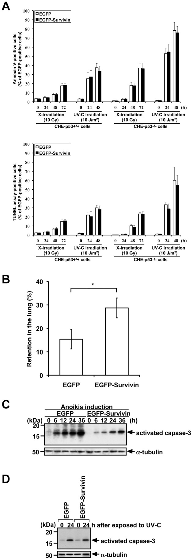
Figure 1. Overexpression of Survivin does not significantly protect radiations-induced apoptosis, but does protect apoptosis under other stresses.

A. Frequency of apoptosis in CHE cells (with p53+/+ and p53−/−) transfected with pEGFP-empty and pEGFP-Survivin after treatment with X-irradiation (10 Gy) and UV-C (10 J/m2). The transfected cells were exposed to IR or UV-C at 24 h after transfection. Transfection frequencies were 80–90%, and EGFP-positive cells were counted for Annexin V-positive or -negative cells (upper panel) and TUNEL assay-positive or -negative cells (lower panel). X-irradiated CHE-p53−/− cells had significantly greater fraction of apoptosis-positive cells when compared to X-irradiated CHE-p53+/+ cells (P<0.03 for Annexin V staining and P<0.08 for TUNEL assay), and UV-C irradiated CHE-p53−/− cells had significantly greater fraction of apoptosis-positive cells when compared to UV-C irradiated CHE-p53+/+ cells (P<0.001 for Annexin V staining and P<0.02 for TUNEL assay). Apoptosis-positive cells were not significantly decreased in pEGFP-Survivin-transfected cells when compared to pEGFP-transfected cells in each case (P>0.4 for Annexin V staining and P>0.4 for TUNEL assay). Values indicate means ± S.D. (n = 3). B. Increase of the retention in the lung after intravenous injection of EGFP- or EGFP-Survivin-expressing cells. The number of surviving CHE-p53−/− cells in the lung was significantly increased when compared to the number of surviving control cells expressing EGFP. Values indicate means ± S.D. of six mice. *Significant difference (P<0.005). C. Caspase-3 activation in CHE-p53−/− cells transfected with pEGFP-empty and pEGFP-Survivin after anoikis induction. The transfected cells were detached from extracellular matrix and simultaneously serum-starved to induce anoikis at 24 h after transfection. Cells were suspended in serum-free medium for 6–36 h, harvested, and lysed in Laemmli SDS-sample buffer for immunoblot analysis with anti-activated caspase-3 antibody. Transfection frequencies were checked by using fluorescence microscopy and confirmed to be 80–90%. D. Caspase-3 activation in CHE-p53−/− cells transfected with pEGFP-empty and pEGFP-Survivin after treatment with UV-C (10 J/m2). The transfected cells were exposed to UV-C at 24 h after transfection. Cells were cultured for 24 h, harvested, and lysed in Laemmli SDS-sample buffer for immunoblot analysis with anti-activated caspase-3 antibody. Transfection frequencies were checked by using fluorescence microscopy and confirmed to be 80–90%.
