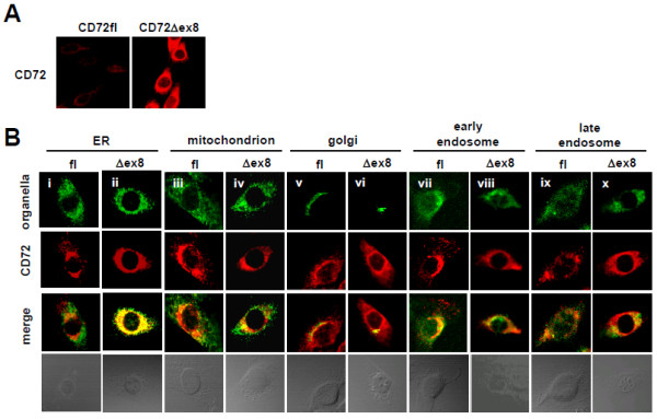
Figure 5.

CD72Δex8 is accumulated in ER. A: Intracellular staining CD72 in Balb/c-3T3 transfectants. B: Intracellular localization of CD72fl and CD72Δex8 in Balb/c-3T3 transfectants. Because of the relatively low fluorescence of CD72fl, its fluorescence intensity was amplified. Proteins localized in ER (i, ii), mitochondria (iii, iv), Golgi apparatus (v, vi), early endosomes (vii, viii), and late endosomes (ix, x) are shown in green. CD72fl and CD72Δex8 are shown in red. Cells were observed by confocal microscopy. Representative data from five experiments are shown.
