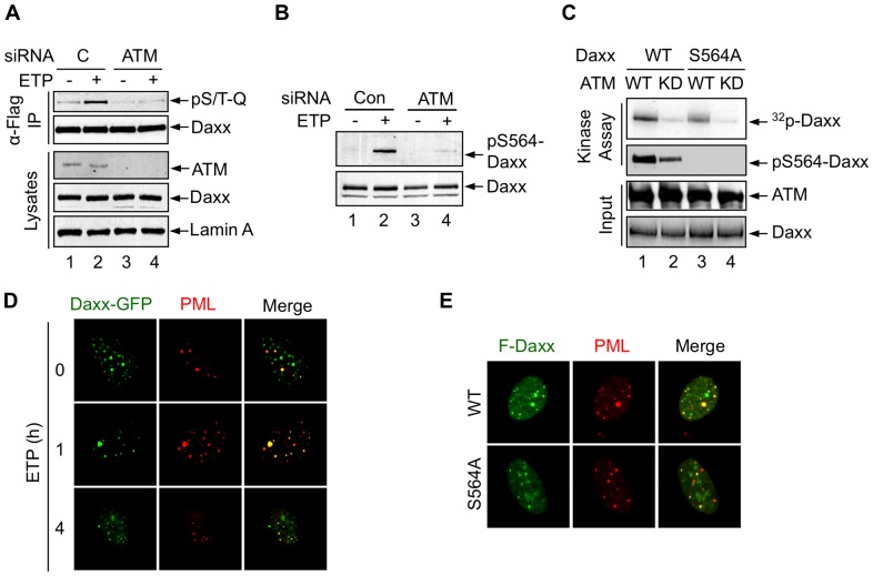
Figure 3. Daxx is phosphorylated by ATM in vivo and in vitro.
(A) H1299 cells treated with a control (C) siRNA or ATM siRNA were transfected with Flag-Daxx and treated with etoposide. Cell lysates and immunoprecipitated Flag-Daxx were examined by western blot using anti-ATM, lamin A, anti-Daxx, and anti-pS/T-Q antibodies. (B) Phosphorylation of endogenous Daxx upon DNA damage in H1299 cells treated with ATM siRNA or control siRNA. (C) ATM phosphorylates Daxx at Ser564 in vitro. Top two panels: phosphorylated Daxx was detected by autoradiography (32P-Daxx) and western blot (pS564-Daxx). Bottom two panels: input of ATM and Daxx proteins were analyzed by western blot and Coomassie Blue staining, respectively. (D) H1299 cells transfected with the GFP-Daxx were untreated (0) or treated with ETP for 1 or 4 h. Endogenous PML was detected by anti-PML antibody and Texas Red-labeled secondary antibody. Images were captured using confocal microscopy. (E) H1299 cells were transfected with Flag-Daxx or Flag-Daxx S564A. Cells were stained with anti-Flag and anti-PML antibodies.

