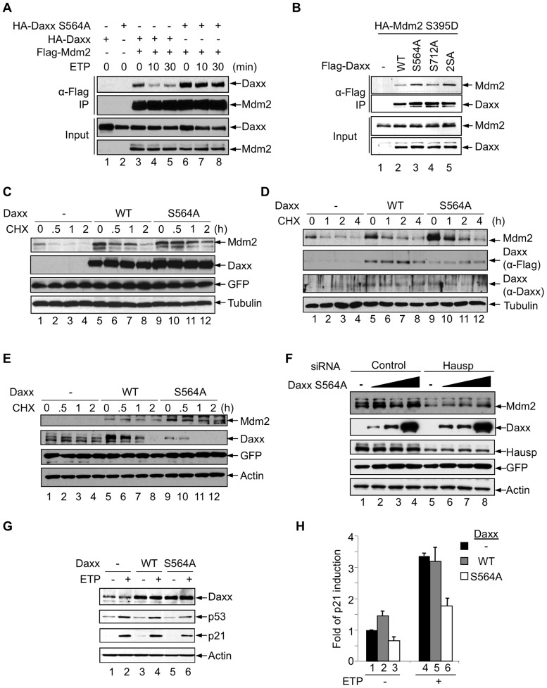
Figure 4. Phosphorylation of Daxx at Ser564 regulates its interaction with Mdm2.
(A) Daxx S564A has enhanced binding to Mdm2 upon DNA damage. H1299 cells were transfected with either HA-Daxx or HA-Daxx S564A alone, or together with Flag-Mdm2. Cells were treated with MG-132 (20 μM) for 4 h and ETP (20 μM) for the indicated times. Cell lysates were incubated with M2 beads. Input and immunoprecipitated proteins were analyzed by western blot. (B) HA-Mdm2 S395D was transfected alone and together with the indicated Flag-tagged Daxx plasmids into p53-/- Mdm2-/- MEFs. Cell lysates were incubated with M2 beads. The input lysates and immunoprecipitated proteins were analyzed by western blotting. (C) H1299 cells were transfected with HA-Mdm2 alone or together with Flag-Daxx or Flag-Daxx S564A. Cells were treated with CHX (50 μg/ml) for the indicated time periods and subjected to western blot analysis. Tubulin and co-transfected GFP are shown as controls for sample loading and transfection efficiency, respectively. (D) Daxx S564A can prolong the half-life of endogenous Mdm2. U2OS cells stably expressing Flag-Daxx or Flag-Daxx S564A were treated with CHX (20 μg/mL) for the indicated time. The expression of Daxx was detected separately by anti-Daxx and anti-Flag antibodies. (E) Daxx S564A can prolong the half-life of Mdm2 upon DNA damage. Flag-Mdm2 was transfected alone or together with the indicated HA-tagged Daxx plasmids into p53-/- Mdm2-/- MEFs. Cells were treated with ETP (20 μM) for 1 h prior to cycloheximide (CHX, 50 μg/ml) treatment for indicated time. GFP and actin are shown as controls for transfection and sample loading, respectively. (F) The effect of Daxx S564A on Mdm2 is dependent upon Hausp. Increasing amounts of Flag-Daxx S564A were transfected into U2OS cells treated with either control or Hausp siRNA and analyzed by western blot. Actin and GFP are shown as controls for sample loading and transfection efficiency, respectively. (G, H) Daxx S564A inhibits p53-mediated gene expression. MCF-7 (G) and U2OS (H) cells stably expressing Flag-Daxx or Flag-Daxx S564A were treated with 10 μM ETP for 8 h. Protein (G) and RNA (H) expression was analyzed by western blot and quantitative RT-PCR, respectively.

