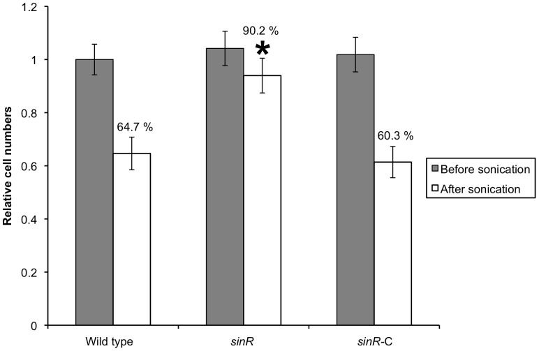
Figure 4. Strength of biofilms formed by P. gingivalis strains.
Standardized cultures of P. gingivalis were inoculated into dGAM in saliva-coated 12-well polystyrene plate and incubated statically at 37°C for 60 h, and the resulting biofilms were sonicated for 1 s. Immediately after sonication, supernatants containing floating cells were removed by aspiration and the biofilm remnants were gently washed with PBS. P. gingivalis genomic DNA was isolated from the biofilms, and the numbers of P. gingivalis cells were determined using real-time PCR. Relative amounts of bacterial cell numbers were calculated based on the number of wild type cells without sonication and assigned a value of 1.0. The percentages shown indicate the amount of remaining biofilm after sonic disruption. Duplicate experiments were independently repeated three times with each strain. Standard error bars are shown. Statistical analysis was performed using Welch's t test. *P<0.001 in comparison with the wild type strain.

