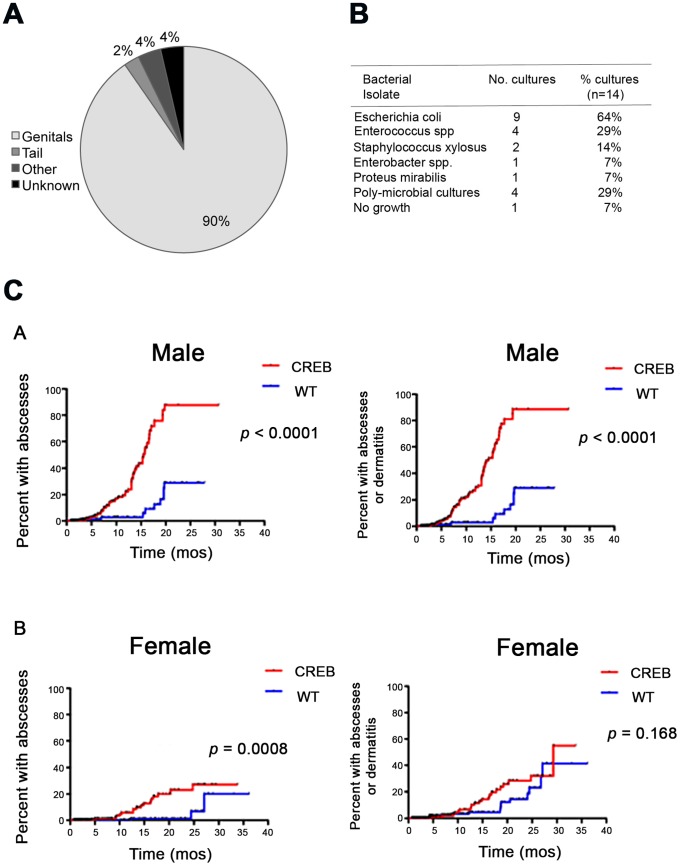
Figure 1. Comparison of abscess location and types of infection.
(A) Numbers of mice that developed skin and soft-tissue abscesses were evaluated and the location of abscesses was recorded. The majority (90%) of skin and soft-tissue abscesses were located in the genital region. The remaining abscesses were discovered on the tail (2%), involved the eyes, kidney, and abdomen (4%), or not recorded (4%). (B) Types of bacteria identified in 14 abscesses cultured from CREB TG mice, the numbers of positive cultures, and percent of each type of bacteria are listed. (C) Estimated Cumulative Incidence of skin infections in CREB TG and WT mice. Kaplan-Meier analysis was performed to take censoring into account in estimating the cumulative risk with age of developing a first skin abscess or non-abscess dermatitis in CREB TG and WT, if male and female mice could be followed indefinitely under breeding colony conditions. (A) CREB TG males developed skin abscesses alone earlier and with a higher overall risk than WT male mice (p<0.0001 by log-rank test). Similarly, CREB TG male mice were at greater risk of developing abscesses or non-abscess dermatitis than WT mice (p<0.0001). (B) CREB TG female mice developed skin abscesses earlier than WT female mice (p = 0.0008). However, the time to first appearance of abscess or non-abscess dermatitis did not appear significantly different between CREB TG female and WT female mice (p = 0.168).

