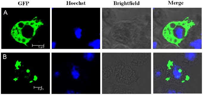
Figure 7. Subcellular localizations of LvSARM in Drosophila S2 cells.

Drosophila S2 cells were transfected with plasmid pAc5.1-LvSARM-GFP. Nuclei were visualized with Hoechst (blue). (A) The LvSARM-GFP fusion protein was widely distributed in the cytoplasm of Drosophila S2 cells, as revealed by confocal microscopy. (B) Drosophila S2 cells transfected with pAc5.1-LvIMD-GFP (LvIMD Accession no. ACL37048) were used as controls.
