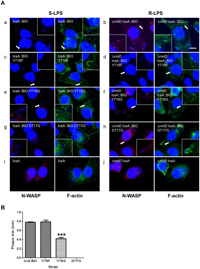
Figure 3. Effect of mutagenesis of IcsA::BIO Y716 and IcsA::BIO D717 on N-WASP recruitment and intercellular spreading.
(A) HeLa cells were infected with mid-exponential phase S. flexneri ΔicsA (S-LPS) or S. flexneri ΔicsA ΔrmlD (R-LPS) having an empty vector or expressing IcsA::BIO, IcsA::BIO Y716F, IcsA::BIO Y716G or IcsA::BIO D717G, and then formalin fixed. HeLa cells and bacteria nuclei were labelled with DAPI (blue), F-actin was labelled with Alexa Fluor 488-phalloidin (green), and N-WASP was labelled with anti-N-WASP antibody and Alexa Fluor 594-conjugated donkey anti-rabbit antibody (red) as detailed in Materials and Methods. IF images were observed at 100×magnification. Arrows indicate N-WASP recruitment and F-actin comet tail formation. Enlargements of relevant region shown for clarity. Strains were assessed in two independent experiments. Scale bar = 10 µm. (B) Plaque assay of S. flexneri ΔicsA strains expressing IcsA::BIO, IcsA::BIO Y716F, IcsA::BIO Y716G or IcsA::BIO D717G. Confluent HeLa cell monolayers were infected with mid-exponential phase S. flexneri strains for 2 h, and plaques were observed 48 h post-infection as detailed in Materials and Methods. 30 plaques were measured from each experiment. Data are represented as mean ± SEM of three independent experiments. ***, P<0.001 (determined by Student’s unpaired one-tailed t test).

