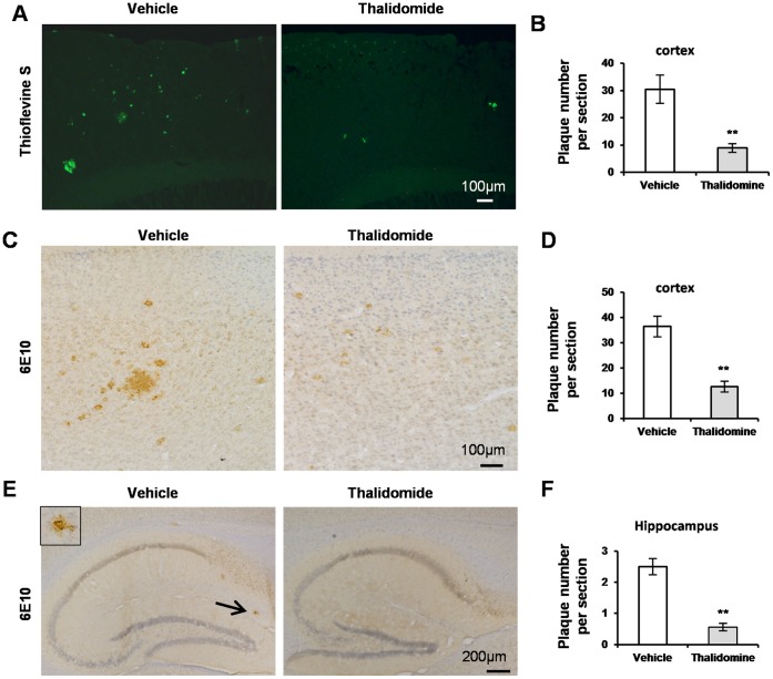
Figure 2. Thalidomide meliorates Aβ pathology.
Representative images showed positive structures of thioflavine S staining, which confirms insoluble Aβ deposits, in the neocortex of 12-month-old APP23 mice with or without thalidomide application for 3 months (A). Thioflavine-positive plaques were counted and statistical analysis showed a significant decrease in the thioflavine-positive number of the neocortex along with thalidomide application vs vehicle group (Mean ± SD, **p<0.01, Student t-test, n = 10 each group) (B). Microphotographic images presented senile plaques which were confirmed by immunostaining of antibody against Aβ1-17 (Clone: 6E10) in the neocortex (C) and hippocampus (E). Counter staining was performed by haemotaxylin. Insert in (E) showed an amplified 6E10-positive plaque pointed by the arrow. Statistical analysis demonstrated a significant decrease in the number of 6E10-positive plaques in the neocortex (D) and hippocampus (F) (Mean ± SD, **p<0.01, Student t-test, n = 10 each group) following thalidomide administration. Bar: 100 µm (A, C), 200 µm (E).

