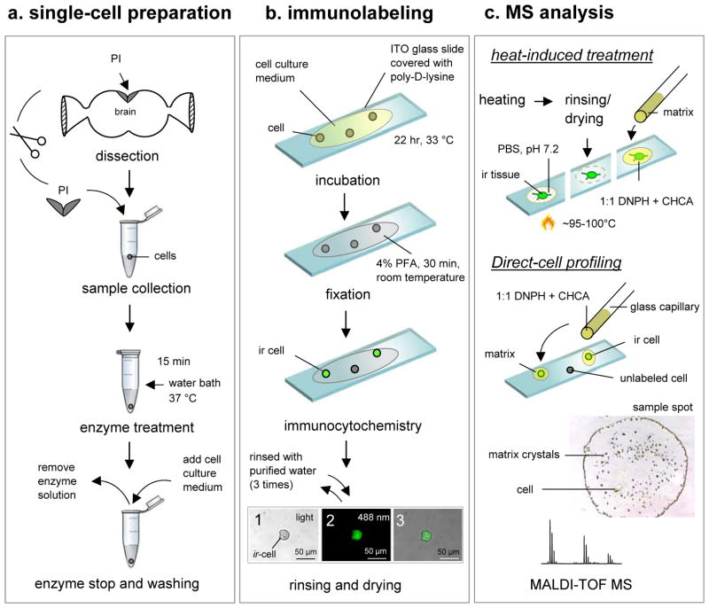
Figure 1. Strategy for ICC-guided peptidomics of individual cells.
The strategy has three experimental stages: (a) single-cell preparation (isolation and culturing), (b) immunolabeling, and (c) individual cell and tissue MS-based characterization, including peptide profiling and identification. Abundant peptides that were observed in the mass spectra are subsequently fragmented by tandem mass spectrometry to reveal the amino acid sequence. The three images in panel (b) show (1) a single RFamide ir PI cell with a cell size of 30 μm under ambient light condition, (2) the same cell as in (a) visualized with a fluorescence microscopy, and (3) merged image of (1) and (2). Scale bar = 50 μm.

