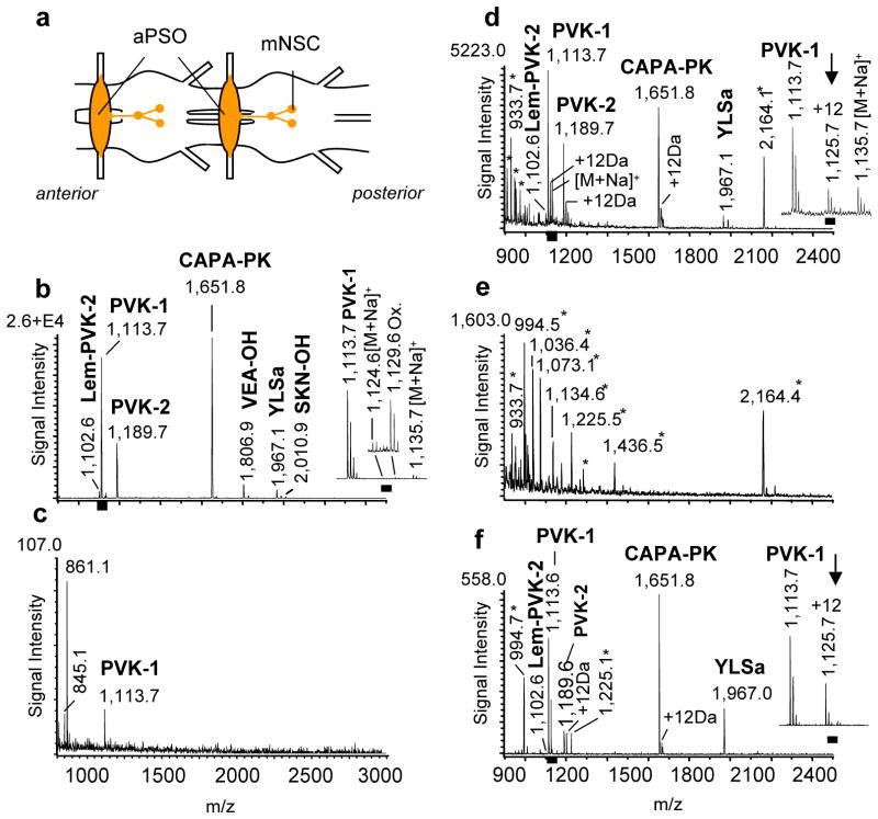
Figure 2. MS analysis of peptides from immunolabeled tissues.
Comparative MS peptidomic analyses of freshly isolated and paraformaldehyde-fixed immunolabeled (PFIL) tissues demonstrated that endogenous peptides can be successfully detected in both types of samples. PFIL samples needed to be treated with high temperature (for 42 weeks old samples) before matrix application or a 2,4-DNPH/CHCA mixture (for samples up to 22 weeks) in order to detect peptides. (a) Simplified schematic of a region of the abdominal ventral nerve cord (aVNC) of P. americana. Peptides are produced in median neurosecretory cells (mNSC) in each abdominal ganglion and transported for storage and release to the abdominal perisympathetic organs (aPSO). Scale bar: 500 μm (b) Mass spectrum of freshly prepared aPSO (MALDI matrix solution contains saturated CHCA solution diluted in a 2:1 ratio with 50 % MeOH/water) (n > 100); Scale bar: 100 μm; (c) Mass spectrum of PFIL aPSO using only CHCA as the MALDI matrix; (n = 20); (d) Representative mass spectrum of a 1 to 22 weeks old PFIL aPSO stored in purified water at 4 °C and treated with the 2,4-DNPH/CHCA MALDI matrix solution (2,4-DNPH dissolved in 70% acetonitrile/water, 0.5% TFA mixed with saturated CHCA dissolved in 60% methanol/water, 0.01% TFA in a 1:1 ratio); (n = 15). (e) Mass spectrum of PFIL-treated aPSO stored for 42 weeks in purified water at 4 °C and treated with the same 2,4-DNPH/CHCA matrix used in (d). No known peptide ion signals were detected in any of the preparations (n = 3). (f) Mass spectrum of a 42 weeks old PFIL aPSO from that VNC which provided also the PFIL aPSO for MS analysis shown in (e), however, that sample was heat-treated (2–5 min at 95–100 °C in PBS, ph 7.2) before 2,4-DNPH/CHCA matrix mixture application. Labeled signals correspond to peptides from the putative capa-gene of P. americana. CAPA-PVKs (Pea-PVK-1 [M+H]+ 1,113.62, Pea-PVK-2 [M+H]+ 1,189.63, Lem-PVK-2 [M+H]+ 1,102.60), CAPA-pyrokinin (CAPA-PK, [M+H]+ 1,651.76) and Pea-YLSamide ([M+H]+ 1,967.00; n = 5. Ion signals marked with an asterisk are unspecific and could be only detected from ir tissues (see also supplemental Figure 1).

