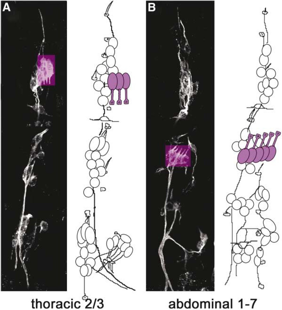
Figure 1. Sense Organs in a Late Embryo.
Thoracic (A) and abdominal (B) segments in the epidermis of an embryo stained with mAb22C10 to label all sensory neurons. Confocal projections are shown on the left in each panel, and schematic drawings on the right show locations of cell bodies. Chordotonal organ neurons are highlighted in purple. The clusters of three thoracic Cho neurons in T2 and T3 (dCh3) are located dorsally and their dendrites point downward, whereas clusters of five Cho neurons in abdominal segments 1–7 (lCh5) are located laterally, and their dendrites point upward. Dorsal is up and anterior is left in all panels. Drawings adapted with permission from Campos-Ortega and Hartenstein [25].

