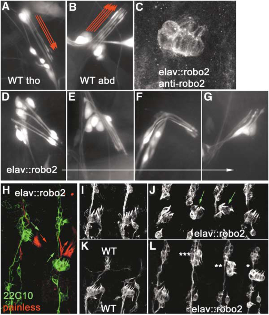
Figure 2. Overexpression of Robo2 in Chordotonal Neurons Transforms Abdominal Organs to a Thoracic Morphology and Position.
(A and B) Wild-type third instar larval thoracic dCh3 and abdominal lCh5 neuron clusters, respectively, expressing GFP from the elav driver. Note the downward-pointing (ventral) orientation of thoracic dCh3 dendrites and the upward-pointing (dorsal) orientation of abdominal lCh5 dendrites (red arrows).
(C) elav::robo2 embryonic lCh5 stained for Robo2.
(D–G) Larval abdominal lCh5s transformed to varying degrees toward the thoracic morphology, with dendrites either pointing downward as in a wild-type thoracic dCh3 (D and E) or twisted in an intermediate position (F) or simply stunted (G).
(H) Sense organs of an elav::robo2 embryo labeled with mAb 22C10 (green) and anti-Painless (red) to show orientation of neurons in two abdominal lCh5s (green arrows), one transformed toward thoracic type (left) and one not (right). Anti-Painless labels the Cho cap cells (red arrows), which do not express Robo2, but follow the orientation of the Cho neurons that do.
(I and K) Wild-type abdominal segments at late stage 14 and stage 15, respectively, stained with 22C10 to show orientation of Cho neurons.
(J) elav::robo2 abdominal lCh5s at stage 14, the earliest stage at which transformed lCh5s (green arrows) can be unambiguously recognized in a fixed preparation.
(L) elav::robo2 abdominal lCh5s at stage 15, showing various classifications of transformation: complete (triple star), partial (double star), and shifted (single star).

