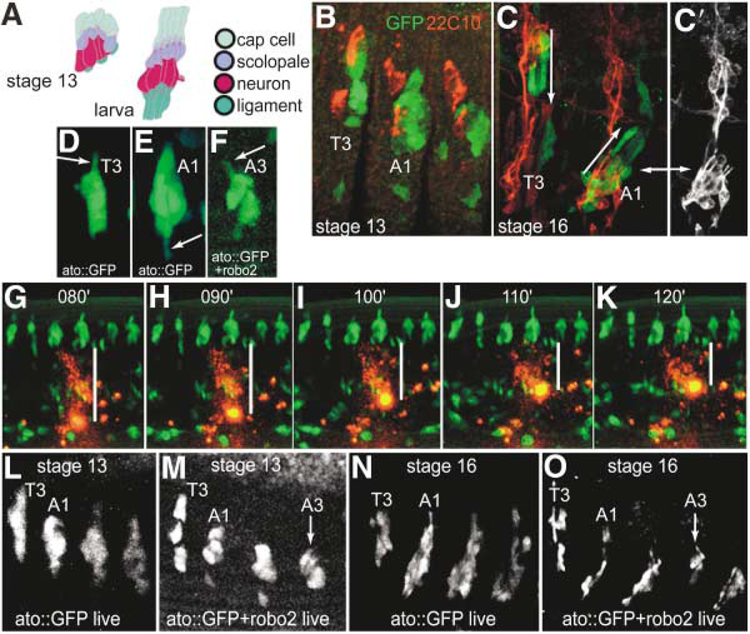
Figure 3. Live Observation of Migrating Chordotonal Organ Precursors.
(A) Schematic representations of abdominal chordotonal organs in a stage 13 embryo versus the larva. Each organ, which is present in abdominal segments as clusters of five (shown), consists of a cap cell, which anchors the organ to the epidermis at the dorsal tip, a scolopale, which ensheathes the dendrite, a neuron, which possesses a prominent dendrite, and a ligament cell, which anchors the organ at its ventral side. This arrangement is reversed in thoracic segments, so that the cap cells point ventrally rather than dorsally (adapted with permission from Campos-Ortega and Hartenstein[25], Figure 9.2).
(B) A stage 13 embryo expressing atonal-GAL4 driven GFP in all Cho precursors (green) and labeled for 22C10 (red), which labels the neurons of all sense organs. T3, third thoracic segment; A1, first abdominal segment in all panels.
(C) A stage 16 embryo labeled in the same manner, showing further refinement of GFP expression such that the orientation of Cho organs is apparent. Cho organs have dendrites pointing ventrally in thoracic segments and dorsally in abdominal segments (arrows). (C′) 22C10 expression alone shows the location of neurons, by which the orientation of the organ can easily be recognized, in relation to the GFP pattern (double arrow points to the same lateral Cho cluster in each panel).
(D–F) Stills from time-lapse series of developing Cho organs in a control embryo (ato::GFP), and an embryo expressing both GFP and Robo2 in Cho organs (see Movies 1 and 2 online). Here, at early stage 14, as Cho organs begin to actively migrate, processes (arrows) can be seen protruding from either the dorsal side in a thoracic segment (D) or the ventral side in an abdominal segment (E) of a normal embryo. In the embryo expressing Robo2 in Cho precursors (F), a process extends from the dorsal side (arrow) of an abdominal Cho precursor, similar to that seen in a thoracic segment.
(G–K) Still images from time-lapse movie of a wild-type (ato::GFP) embryo injected with a spot of DiI to show movement of abdominal Chos relative to the surrounding epidermis. White bars show that the distance from the ventral-most extent of the Cho organs to the center of the DiI spot diminished over time (numbers represent frames of the movie, taken every 1 min).
(L–O) Still images from time-lapse movies of developing control Chos (ato::GFP) versus Robo2-overexpressing Chos (ato::GFP+ robo2) at stages 13 and 16, showing one thoracic segment and three abdominal segments in each panel. Note that by stage 16, the final orientation of the organ is evident, with abdominal Chos positioned more ventrally and oriented oppositely to thoracic Chos (compare [N] to [C]). They reach this position from more equivalent dorsoventral positions at stages 12 and 13 (as in [L] and [M]) by a ventral migration (G–K, and see Movie 1). Note that the A3 lCh5 in the Robo2-overexpressing embryo (O) has failed to migrate and remained in a thoracic position (arrow). The slight dorsal displacement of anterior (T3 and A1) relative to posterior (A2 and A3) Chos at stage 13 (L and M) is caused by the curvature of the germ-band during gastrulation. Less GFP is evident in the ato::GFP+ robo2 shown in (F), (M), and (O) because it contains only one copy of UAS-GFP.

