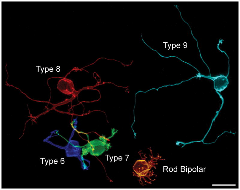
Figure 1.

Five single ON-cone bipolar cells viewed en face in retinal wholemount preparations, illustrating the breadth of dendritic morphology in the outer plexiform layer (OPL). All but the Type 9 cell had been labeled in the same retina, and show their true spatial relationships to one another. The Type 9 cell is from another wholemounted retina, positioned aside these other four cells for direct comparison. Each cell had been labeled with DiI, reconstructed, and then pseudo-colored to discriminate them. Calibration bar = 10 μm.
