
Figure 6.
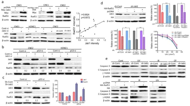
Jab1 associates with Rad51 and contributes to cisplatin (CP), IR and UV resistance. (a) NPC cells were transfected with Myc-Jab1 plasmid DNA or siRNA or transduced with Jab1 adenovirus in the presence or absence of doxycycline (Dox) for 48 h. Cell lysates were prepared 48 h after transfection or from cells stably expressing control (sh-Cont) or Jab1 shRNA (sh-Jab1) and western blotting was performed with the Jab1 and Rad51 antibodies. (Right) Correlation plot of Jab1 and Rad51 blot intensities obtained from control and treated cells. A fair linear correlation was obtained. (b) NPC cells were transfected with siRNA for 48 h, cells lysates were prepared and the levels of Jab1, p53 and Rad51 were determined by western blotting. (c) sh-Cont or sh-Jab1 stable CNE2 cells were transfected with siRNA for 48 h, then total protein and RNA was extracted and expression protein or RNA levels of the Jab1 and p53 and Rad51 were quantified by western blotting (left) or real time qRT-PCR (right). (d) CNE2 cells stably expressing sh-Jab1 were transfected with pcDNA or HA-Rad51 plasmid DNA. (Left) Western blot analyses demonstrated the effective knockdown and ectopic expression of Jab1. (Right and bottom) Colonies were stained with crystal violet 10 days after CP, IR and UV exposure. (e) (Top) CNE2 cells stably expressing sh-Cont or sh-Jab1 were exposed to UV or IR or CP. (Bottom) Cells stably expressing sh-Jab1 were transfected with pcDNA or HA-Rad51 plasmid DNA and then exposed to CP, IR and UV. Cleaved caspase-3 and γ̃H2AX were examined 48 h after exposure. All data represent three independent experiments, mean±s.d. *P <0.05, **P <0.01.