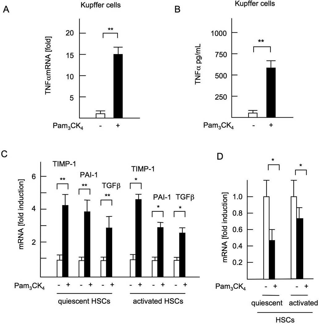
Figure 3. A synthetic TLR2 ligand activates both Kupffer cells and HSCs.
WT Kupffer cells and HSCs were isolated and cultured in the presence of 5 µg/ml Pam3CK4. (A) mRNA expression of TNFα in Kupffer cells. (B) TNFα concentrations in the supernatant from WT Kupffer cell-culture. (C) mRNA expression of fibrogenic genes in quiescent HSCs and culture-activated HSCs. (D) Bambi mRNA expression in HSCs. Genes were normalized to 18S RNA as an internal control. Data represent mean ±SD, *p<0.05. **p<0.01. n.s.; not significant.

