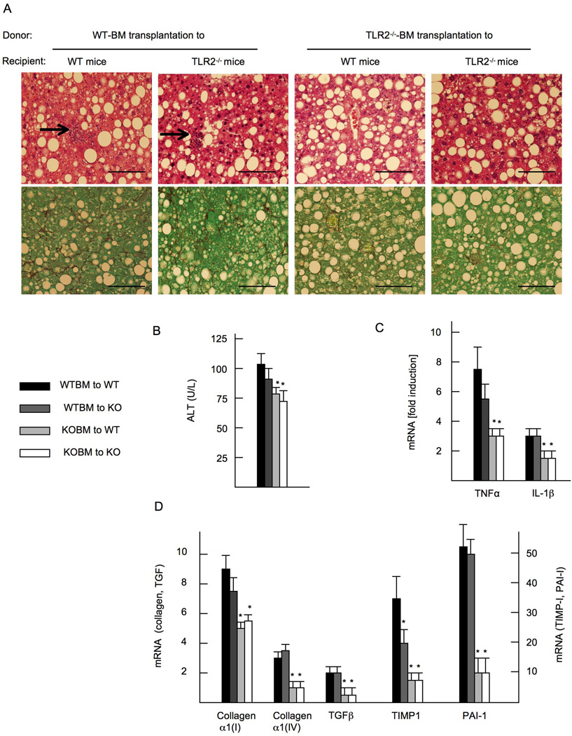
Figure 4. Hematopoietic cells including Kupffer cells are crucial for the development of liver inflammation and fibrosis in NASH.
TLR2 bone marrow (BM) chimeric mice, in which hepatic macrophages were reconstituted with transplanted BM cells, were generated. Mice on CDAA diet are presented. (A) HE staining (upper) and Sirius red staining (lower). WT BM-transplanted mice show inflammatory cell infiltration (arrows), which are blunted in TLR2−/− BM transplanted mice. Liver fibrosis is attenuated in TLR2−/− BM transplanted mice. Original magnification, × 400. Bar 100 µm. (B) Serum ALT levels. (C) mRNA expression of TNFα and IL-1β. (D) mRNA expression of fibrogenic genes. Genes were normalized to 18S RNA as an internal control. Data represent mean ±SD, *p<0.05.

