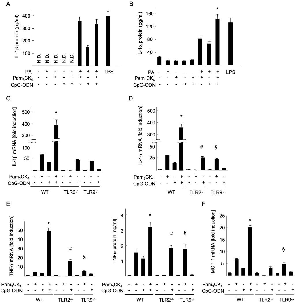
Figure 7. TLR2 and TLR9 ligands synergistically produce inflammatory cytokines in Kupffer cells.
Kupffer cells were isolated from WT, TLR2−/− and TLR9−/− mice, and cultured in the presence of 5 µg/ml Pam3CK4, 5 µg/ml CpG-ODN and/or 200 µM palmitic acid. (A,B,E) IL-1β (A), IL-1α (B) and TNFα (E, right) proteins in the supernatant. (C-F) mRNA expression of IL-1β (C), IL-1α (D), TNFα (E, left) and MCP-1 (F). Genes were normalized to 18S RNA as an internal control. N.D; not detected. Data represent mean ±SD, *; significant differences to WT KCs treated with Pam3CK4 or CpG-ODN, #; significant differences to WT KCs treated with CpG-DNA, §; no significant difference with WT KCs treated with Pam3CK4.

