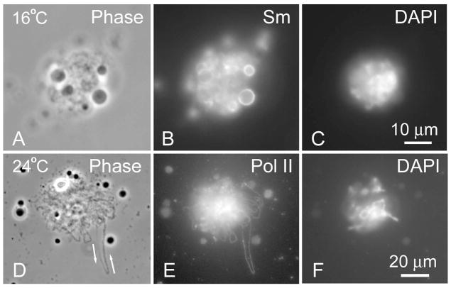
Fig. 2.
Effect of temperature on expansion of sperm heads. a–c A single sperm head 44 h after injection showed minimal expansion when the recipient oocyte was held at 16°C. Note the associated nuclear bodies with a relatively unstained core and a surrounding shell that stains strongly with mAb Y12 against the “Sm” epitope (symmetric dimethylarginine). d–f A single sperm head 42 h after injection was more expanded when the recipient oocyte was held at 24°C. One very long transcription loop extends out from the central cluster. The phase contrast image (a) shows the characteristic “thin-to-thick” loop matrix of ribonucleoprotein, which indicates the direction of transcription (arrows). The pol II antibody (b) shows uniform staining along the entire loop, presumably due to close packing of pol II molecules on the DNA axis. The axis itself is not detectable by DAPI staining (c) because of its extreme attenuation.

