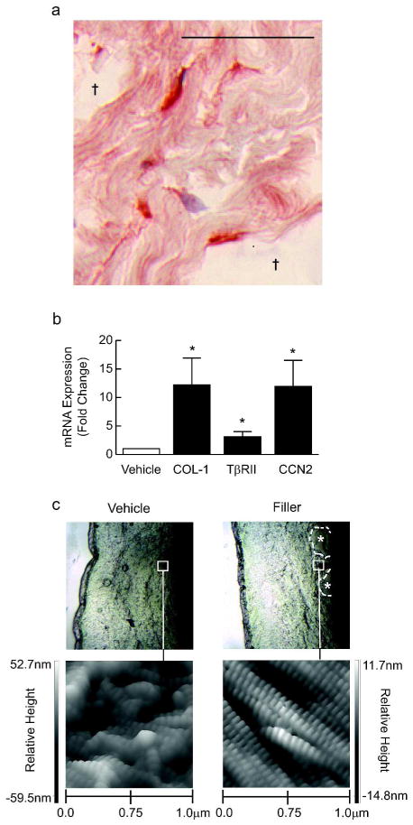
Figure 2. Enhanced structural support up-regulates TGF-β pathway and collagen deposition in aged human skin fibroblasts.
Skin was obtained 4 weeks after injection of vehicle or filler. (A) Image of pools of injected filler (†), with adjacent elongated fibroblasts immunostained for type I procollagen (bar=50μm). (B) Fibroblasts from vehicle- and filler-injected skin were isolated by laser capture microdissection, and analyzed for type I procollagen (COL-1, n=9), type II TGF-β receptor (TβRII, n=9) and connective tissue growth factor (CCN2, n=7). Means+SEM, *p<0.05. (C) Nanoscale structure of collagen fibrils imaged by atomic force microscopy (n=3). Upper panels display probe location in mid dermis. Collagen fibrils with characteristic banded pattern appear intact, tightly packed, and spatially organized in filler-injected skin, but fragmented and disorganized in vehicle-injected skin.

