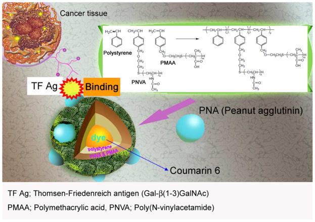
Figure 1.
Overall design of the nanobeacon for imaging TF-associated colorectal cancer. The probe is comprised of a fluorescent dye, Coumarine 6 encapsulated in the center core by a polystyrene copolymer. The complex was then grafted with PMAA and PNVA polymers on the surface. Here, the PMAA was used as a handle to achieve the covalent binding of PNA molecules, which are the recognition molecules for the TF antigen. PNVA was used to help reduce or obviate nanobeacon interacting with the mucous tissue in the GI tract. The average size of the nanobeacon is approximately 350 nm.

