
Figure 4.
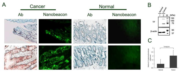
A) The expression of TF antigen on human colonic tissue from normal controls and cancer patients were detected using PNA molecules immobilized on the surface of the nanobeacon and confirmed by immunohistochemistry. Strong fluorescence from representative cancer sections (observation of 60 slides collected from 7 colorectal cancer patients) is correlated to a positive signal in IHC staining of the consecutive slides. Exposure time in fluorescence observation was 400 ms with magnification at 200x each. B) Representative results of Western blot (immunoblot) analysis of TF antigen expression in clinical human tissues with and without colorectal cancer. Beta-actin was shown as a loading control. TF antigen detected in human colorectal tissue samples which molecular weights are approximately 147, 84 and 77 kDa, respectively. C) A relative mRNA expression level of β1,3- galactosyltransferase in clinical human colorectal tissues with and without cancer (n=3) analyzed by qPCR. (* p=0.011).