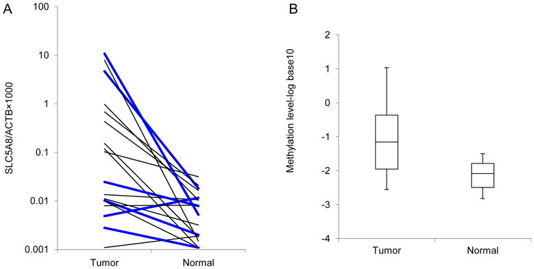
Fig. 3.

Results of QMSP analysis. (A) Methylation levels of SLC5A8 in tumor and adjacent non-tumor tissues from lung cancer patients were measured by QMSP. Calculation of SLC5A8/β-actin ratios was based on the fluorescence emission intensity values for both SLC5A8 and β-actin obtained by quantitative real-time PCR analysis. The relative amount of methylated promoter DNA was higher in tumor compared with adjacent non-tumor tissues (14/23, 60.9%). Results of 5 pairs were excluded because methylation levels of one of these pairs were below the limit of detection. Bold lines represent low SLC5A8 expression in tumors. (B) Distribution of methylation level in two groups is presented. Average methylation level (log10) is −1.16 and −2.09 in tumor and adjacent non-tumor tissues, respectively.
