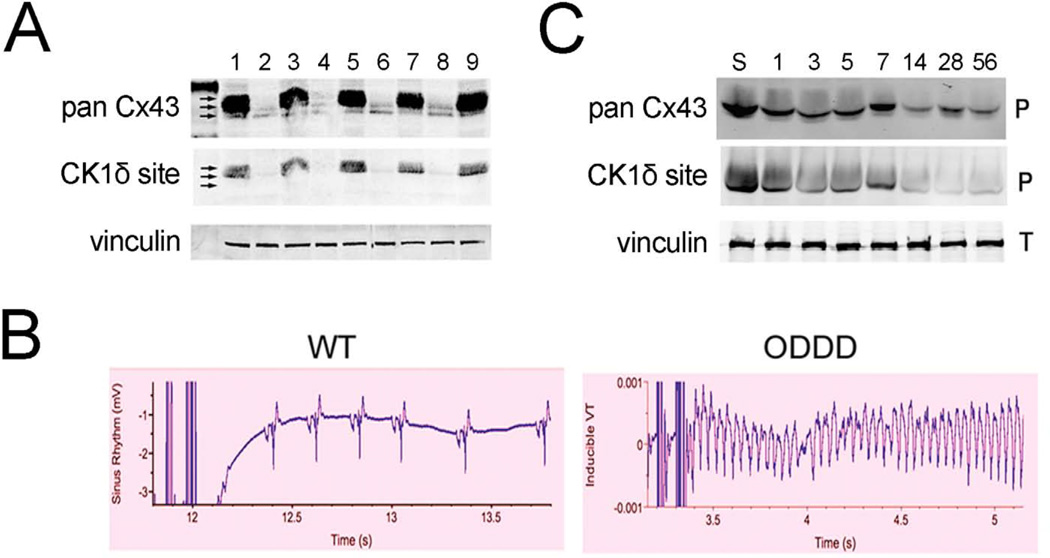
Figure 2. Gap junction remodeling and arrhythmogenesis.
A. Western blot analysis of WT (1, 3, 5, 7, 9) and ODDD mutant (2, 4, 6, 8) hearts showing marked reduction in total Cx43 (pan Cx43) and p325/328/330 phosphoCx43 (CK1δ site). B. Programmed electrical stimulation showing resistance to inducible ventricular tachycardia in WT hearts and induction of sustained ventricular tachycardia in ODDD mutant hearts. C. Western blot analysis of WT hearts subjected to transverse aortic constriction showing progressive reduction in total Cx43 and p325,328/330-Cx43. S is a sham control and numbers refer to days after TAC. Figure adapted from (Kalcheva et al. 2007; Qu et al. 2009).

