
Fig. 1.
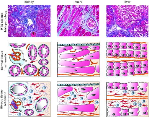
Common features of tissue fibrosis. Macroscopically and microscopically fibrotic organs share obvious commonalities, which has led to the concept of common fibrotic pathways: fibrotic organs are stiff [reflecting excessive extracellular matrix (ECM) deposition], are pale (reflecting rarefication of the vasculature), and have an uneven surface (reflecting fibroblast contraction). Histopathological analysis reveals that tissue fibrosis is unequivocally associated with injury of the parenchyme, accumulation of fibrillar ECM, accumulation of fibroblasts, rarefication of the microvasculature, and a mononuclear infiltrate. The figure illustrates the common appearance of fibrotic kidney, heart, and liver. The photomicrographs display representative MTS-stained sections of fibrotic mouse kidney, heart, and lung. Schematics illustrate the common mechanisms of tissue fibrosis: each organ consists of a functional parenchyme (pink, tubular epithelium in kidney, cardiomyocytes in heart, and hepatocytes in liver) and a connective tissue compartment containing microvessels (orange) and fibroblasts (red, and stellate cells in the liver). Fibrosis is unequivocally associated with expansion of the connective tissue compartment. The fibrotic connective tissue contains ECM fibers (blue), activated fibroblasts (red), and a mononuclear infiltrate (blue). The question is whether common or organ-specific mechanisms lead to this uniform appearance.