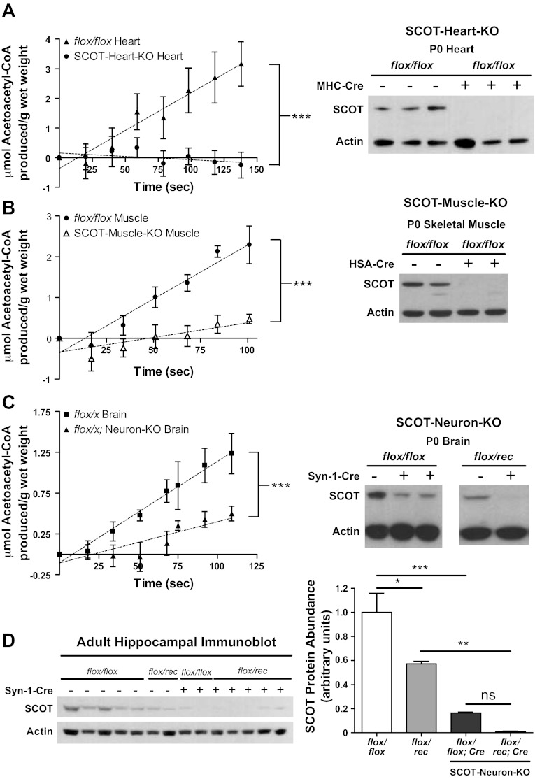
Fig. 3.
Absence of CoA transferase protein and enzymatic activity in tissue-specific SCOT-KO mouse strains. CoA transferase activity was measured spectrophotometrically (left) in tissue lysates derived from heart (A), skeletal muscle (quadriceps/hamstrings) (B), and brains (C) of P0 mice; n = 3–6/group. ***P < 0.001 by linear regression t-test. Brains of SCOT-Neuron-KO mice on both flox/flox and flox/rec genetic backgrounds were analyzed (depicted as flox/x). Immunoblots for CoA transferase (SCOT) and actin (right). D: immunoblot (left) and densitometric quantification (right) of CoA transferase protein abundance, normalized to actin in isolated hippocampi from adult SCOT-Neuron-KO mice; n = 5 for flox/flox mice; n = 2 for flox/rec mice; n = 2 for flox/flox:SCOT-Neuron-KO mice; n = 5 for flox/rec:SCOT-Neuron-KO mice. ***P < 0.001, **P < 0.01, and *P < 0.05 by 1-way ANOVA. ns, Not significant.

