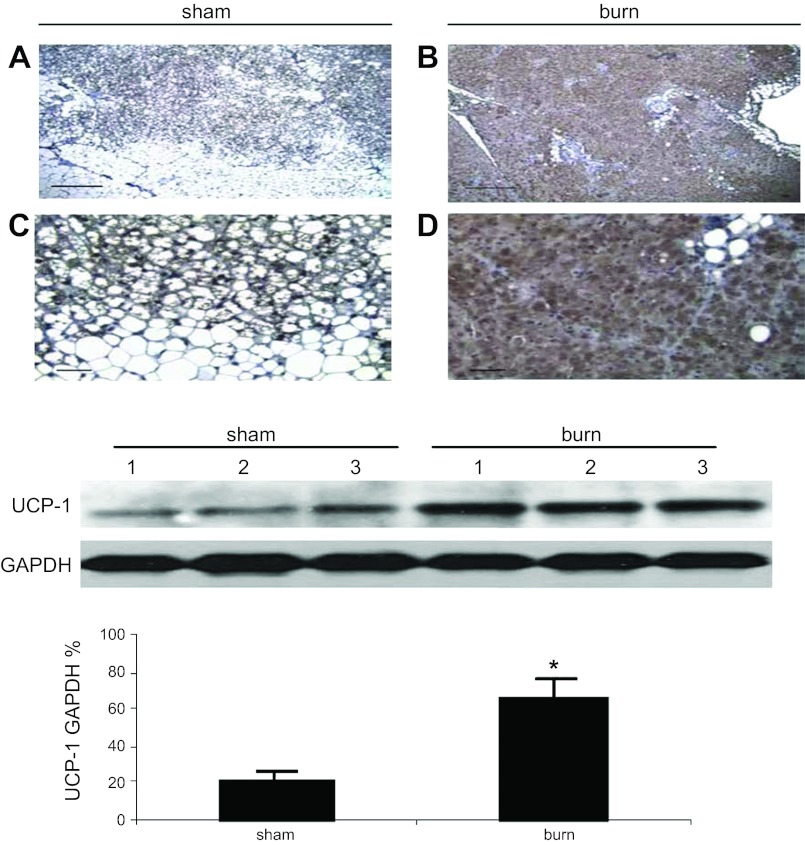
Fig. 5.
Effect of burn injury on uncoupling protein-1 (UCP1) expression in iBAT. A–D: UCP1 expression (brown staining) is observed under low- (×100, A and B) and high- (×400, C and D) power view in sham burn (A and C) and burn animals (B and D). Four slides from 4 sham animals and 5 slides from 5 burned animals were reviewed. Scale bars, 1 mm (A and B); 100 μm (C and D). E: Western blot to identify UCP1 expression in iBAT in animals with or without thermal injury. Results of densitometry are shown in the bar graph. UCP1 expression in burned animals is significantly increased compared with sham burn animals. Denisitometry values are calculated based on mitochondrial loading controls and GAPDH expression. Values are shown as means ± SE in densitometry units (A.U.); n = 6 (sham burn), 6 (burn). UCP1 expression in burned animals was significantly increased compared with sham treated controls *P < 0.01.

