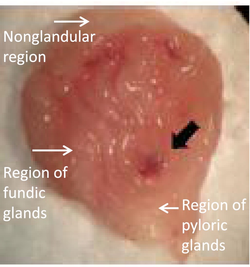
Fig. 3.
Ulcer formation in the glandular region of the stomach in NHE8−/− mice. Stomach from NHE8−/− mice was collected and opened to expose the lumen side. The stomach lumen was rinsed with PBS and then observed under a stereomicroscope (Nikon SMZ-10). Image shown here was a representative photo taken from one NHE8−/− mouse using an Olympus digital camera.

