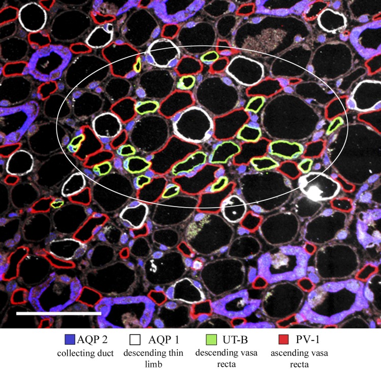
Fig. 7.
Immunolocalization of AQP2, AQP1, UT-B, and PV-1 in transverse section from rat inner medulla, located ∼250 μm below the outer medulla. A single vascular bundle (enclosed by white circle) and associated DTLs lie in the intercluster region. Ascending vasa recta (AVR) nearly always lie between AQP1-positive DTLs and UT-B-positive descending vasa recta (DVR). Scale bar 50 μm.

