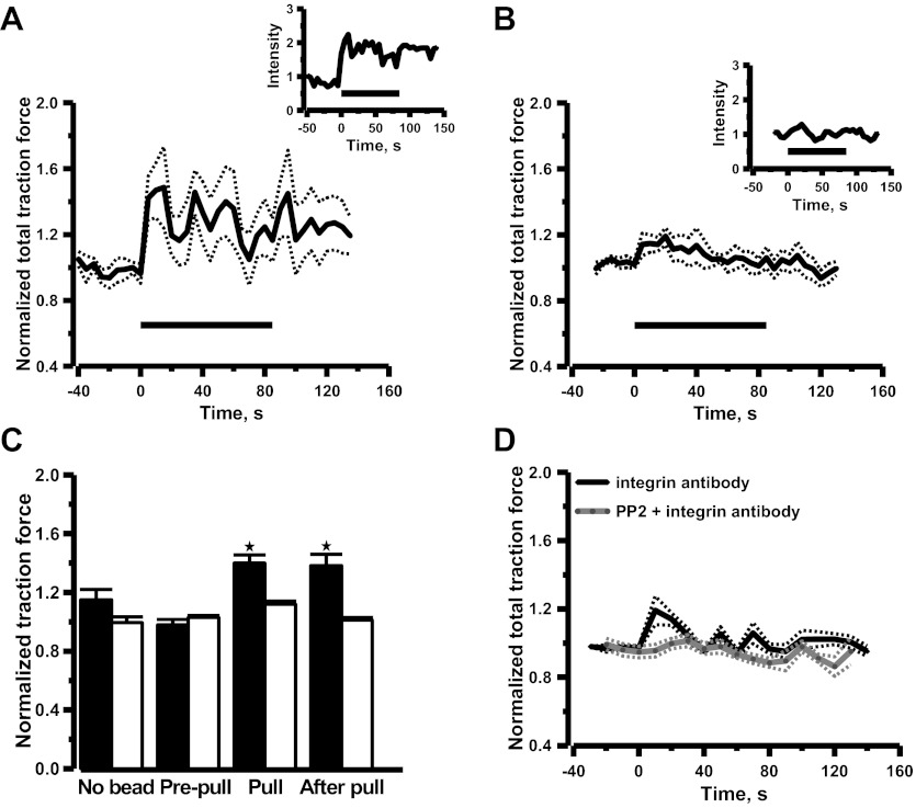
Fig. 3.
Mean time courses of changes in cell traction force induced by pulling paramagnetic beads coated with β1-integrin antibody (A), paramagnetic beads coated with control isotype antibody (B), and soluble β1-integrin antibody alone without paramagnetic beads (D). Time courses from individual cells are shown in insets. Horizontal lines indicate duration of pulling. Dotted lines are ± SE. Cell traction force was normalized with the traction force measured after paramagnetic beads attached (prepull). Mean normalized data are shown in histograms (C). Solid bars and opened bars are β1-integrin antibody and control isotype antibody, respectively. Error bars are SE. *Significant difference compared with the prepull control (P < 0.05, n = 6 cells). The β1-integrin antibody was added to the bath at t = 0 at (D). Preincubation of Src inhibitor PP2 (5 μM) abolished the increase of cell traction force induced by soluble β1-integrin antibody ( n = 6 cells).

