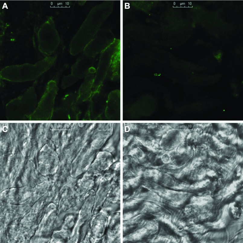
Fig. 7.
Immunofluorescence of activated β1-integrin detected with antibody HUTS-4 in freshly isolated renal VSMCs after activation by antibody Ha2/5 (A) and control isotype antibody (B). Corresponding transmitted light images are shown in C and D, respectively. Strong immunofluorescence of activated β1-integrin was localized on the plasma membrane after activation. Scale bars in C and D = 10 μm. Mean immunofluorescence intensity was increased by 110 ± 8% (3 experiments, 42 cells) after activation compared with the control.

