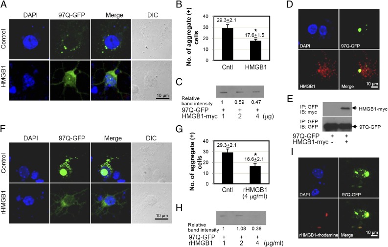
FIGURE 9.
Effect of HMGB1 on the formation of 97Q-GFP aggregates in mouse brain primary striatal cells. (A–E) Mouse embryonic primary striatal neuronal cells were isolated at day 14 and then cotransfected with 97Q-GFP and HMGB1 plasmids. (A) The distribution of 97Q-GFP aggregates was observed under confocal microscopy. (B) The number of aggregate-containing cells was counted among 100 GFP-positive cells. Empty plasmid was used for the control group (Cntl). (C) Filter-trap analysis was performed after cotransfection of 97Q-GFP and HMGB1 plasmids. Colocalization of 97Q-GFP and HMGB1 after cotransfection of both plasmids (D) and immunoprecipitation (IP) analysis (E). (F–H) Mouse embryonic primary striatal neuronal cells were transfected with 97Q-GFP plasmid for 24 h and then treated with rHMGB1 protein at 4 μg/ml. (F) Distribution of 97Q-GFP aggregates was analyzed by confocal microscopy. (G) The number of aggregate-containing cells was counted among 100 GFP-positive cells. Empty plasmid was used as the control group (Cntl). Filter-trap analysis was performed using the cells that were transfected with 97Q-GFP and incubated with rHMGB1 protein. (I) Primary striatal neuronal cells were transfected with 97Q-GFP plasmid, and rhodamine-conjugated rHMGB1 protein was treated. The colocalization of 97Q-GFP with rHMGB1 protein was observed by confocal microscopy. *p < 0.05 (n = 3). DIC, Differential interference contrast; IB, immunoblotting.

