Figure 3.

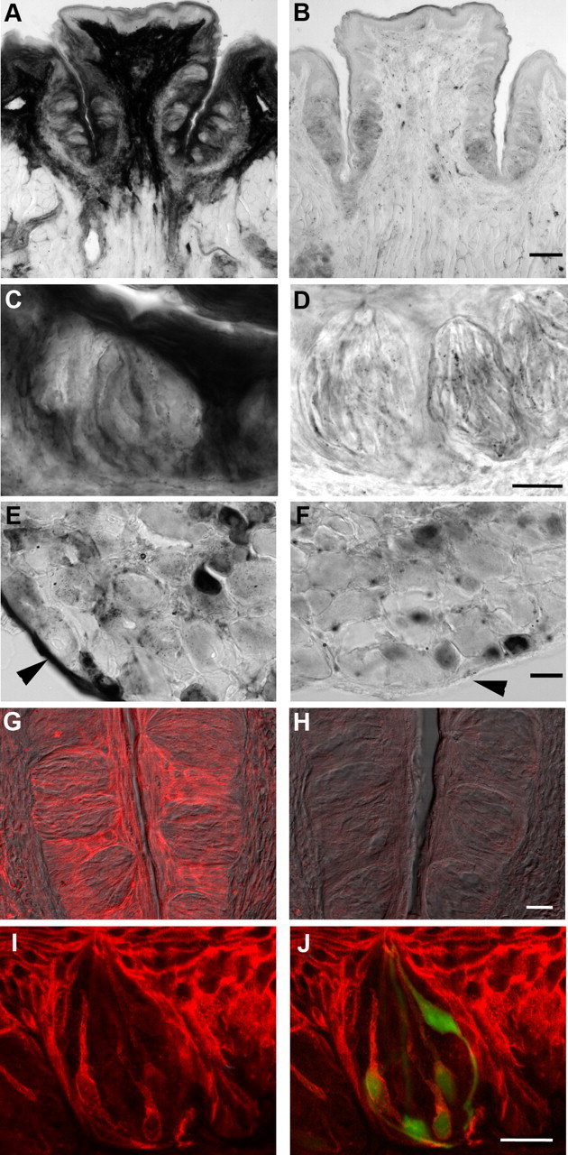
Enzyme histochemical and immunofluorescent localization of ecto-5′-nucleotidase, NT5E. A, Cryosection of wild-type vallate papilla, developed to reveal enzymatic conversion of AMP to adenosine at pH 7.0. Very strong enzymatic activity, seen as dark staining, is present in the connective tissue core of the papilla, in non-taste epithelium on the lingual surface, and in keratinocytes surrounding taste buds. B, Cryosection of vallate papilla from a Nt5e−/− mouse, stained in parallel with that in A. Note the loss of prominent signal in the core of the papilla, in non-taste lingual epithelium, and in keratinocytes surrounding taste buds. C, At higher magnification, taste buds from wild-type mice can be seen to contain NT5E activity, particularly in slender cells. D, Higher magnification of taste buds from Nt5e−/− mouse. No distinct reaction product is seen in cells, although faint residual staining appears throughout the epithelium and taste buds. E, Dorsal root ganglion from wild-type mouse as in A and C shows that small- and medium-sized neurons, and the epineurium (arrowhead) of the ganglion, are stained for NT5E. F, Dorsal root ganglion from the same Nt5e−/− mouse as in B and D above shows that there is no staining in epineurium (arrowhead), but nucleotidase activity remains in small neurons, consistent with previous results (Sowa et al., 2010). G, Immunofluorescence for NT5E in vallate papilla of wild-type mouse, showing pronounced staining in keratinocytes surrounding the taste buds, and more modest staining in limited numbers of slender taste cells. Image is a merge of immunofluorescence (red) and Nomarski differential contrast optics. H, Sections of vallate papilla from Nt5e−/− mouse, stained in parallel and photographed with identical settings as in G. No signal is detected either in epithelium or in taste cells. I, J, Higher magnification of a vallate taste bud from a Gad1-GFP mouse stained for NT5E. The merged view (J) shows that NT5E signal (red) within the taste bud is in slender, GFP-positive cells (i.e., Type III /Presynaptic cells). Note how NT5E immunostaining (I, J) is similar to the shape and incidence of NT5E histochemical reaction product (C). Scale bars: A, D, 100 μm; B, E, 20 μm; C, F, 25 μm; G, H, 20 μm; I, J, 20 μm.
