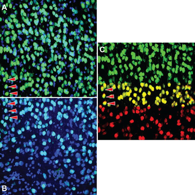
Figure 10.
Illustrating successful registration of images with problematic staining. The nuclear channel is in blue and the neuronal channel in green. The confocal stack in A contains higher background staining when compared to the adjacent stack in B. The arrows indicate the corresponding areas in the two image stacks. C is a crop of the montage of the neuronal channel. It demonstrates accurate alignment using the neuronal channels with neurons from A in green and neurons from B in red. When the two images are well aligned, the neurons in the overlap area correctly appear yellow, as seen here. Images in this figure are from dataset #1.

