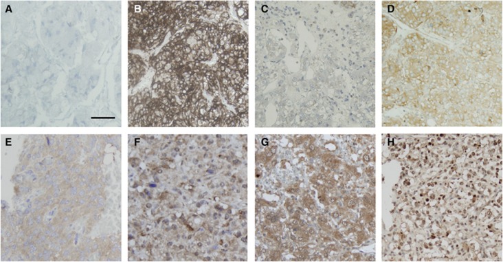
Figure 2.
Expression of the candidate biomarkers in PCC/PGL by immunohistochemistry. Representative sections of tumours showing (A) a negative tumour core . Panel (B) shows strong membranous expression of CaIX in a VHL PCC, not replicated in (C) the matching normal medulla sample. Panel (D) shows a case of positive granular cytoplasmic expression of CaIX, typical of all the non-VHL cases (both sporadic and familial) tested in our series. Panels (E) and (F) are representative of mTor and AKT expression. Samples showing Hif-1α cytoplasmic and nuclear expression are reported in panels G and H, respectively. Magnification × 200, Bar=100 μm.

