Figure 3.
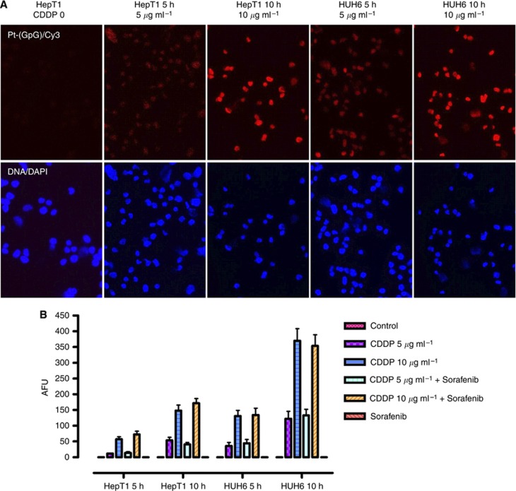
DNA adducts in HB cells treated with CDDP alone and in combination with sorafenib. (A) Formation of Pt-(GpG) adducts was detected by Cy3 immunostaining after incubation of CDDP (5, 10 μg ml−1) for 5 or 10 h. Nuclear staining was performed with DAPI. (B) Adduct levels in the nuclear DNA of HB cells were calculated as fluorescence units (AFUs). AFUs were higher in HuH6 compared with HepT1 cells and were not reduced by the combination with sorafenib. Data represent mean±s.d. from triplicates.

