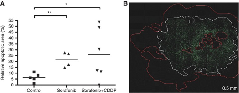
Figure 5.
Apoptotic effect. (A) Relative apoptotic areas are increased in sorafenib- (n=4) and sorafenib/CDDP- (n=6) treated mice compared with control (n=5). Means and relative apoptotic area for each animal are shown (*, **P=0.03, student t-test). (B) TUNEL assay revealed apoptotic cells containing DNA strand nicks as bright green fluorescent cells (white border) within vital cells (red bordered).

