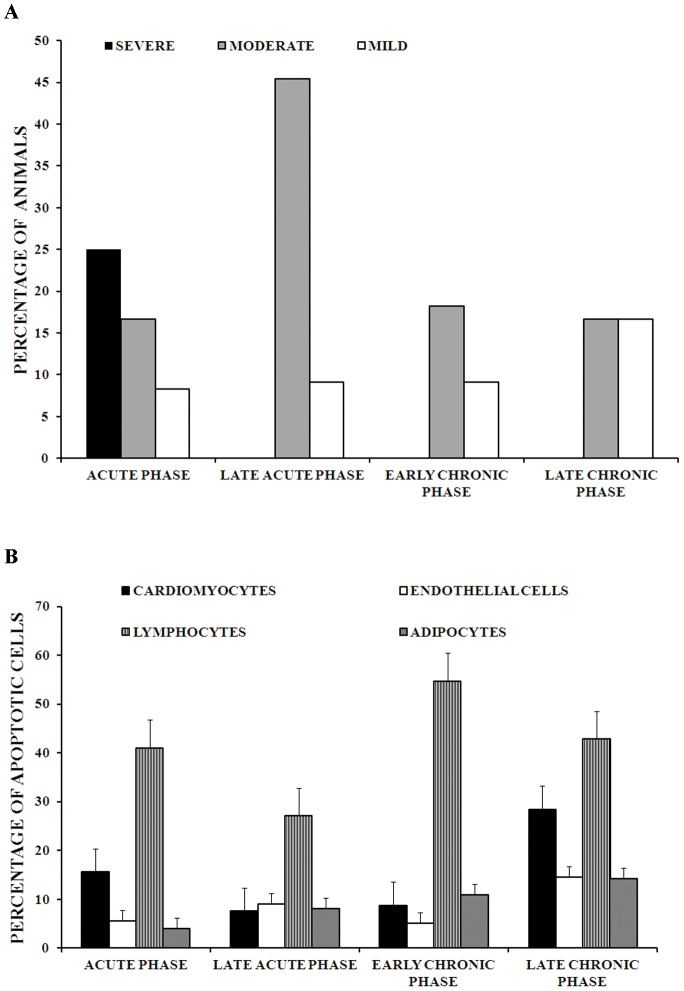
Figure 1. Necrosis and apoptosis during cardiac remodeling in guinea pig infected with T. cruzi.
1A. Degrees of necrosis in cardiac tissue of guinea pig infected with T. cruzi. Degrees of necrosis were determined semiquantitatively: Mild (5%–15% of cardiomyocytes of the entire section), 3. Moderate (16–45% of cardiomyocytes of the entire section) and 4. Severe (more than 45% of cardiomyocytes of the entire section). 1B. Percentages of number of apoptotic cells in cardiomyocytes, lymphocytes, endothelial cells and adipocytes of cardiac tissue of guinea pig infected with T. cruzi. Percentage of cells with apoptosis was determined in 30 microscopy fields. Acute phase: n = 12, late acute phase: n = 11, early chronic phase: n = 11 and chronic phase: n = 6. The bars and error bars represent mean ± standard errors.

