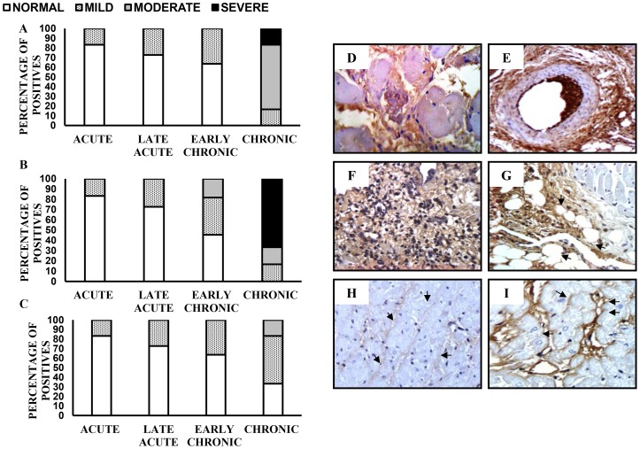
Figure 4. Distribution of collagen isotypes in cardiac tissue of guinea pig infected with T. cruzi.
Deposition of collagen I (A), collagen III (B) and collagen IV (C) during the course of infection. Moderate increase of interstitial collagen I, at 365 dpi, 400× (D). Increase of perivascular collagen I, at 365 dpi, 400× (E). Severe increase of interstitial collagen III and moderate inflammation, at 365 dpi, 400× (F). Increase of collagen III near epicardial adipose tissue at 165 dpi, 400× (G). Detection of collagen IV in non-infected guinea pig (H). Increase of collagen IV in the basement of cardiac fibers, at 365 dpi, 400× (I). Acute phase (20–25 dpi): n = 12, late acute phase (40–55 dpi): n = 11, early chronic phase (115–165 dpi): n = 11 and chronic phase (365 dpi): n = 6.

