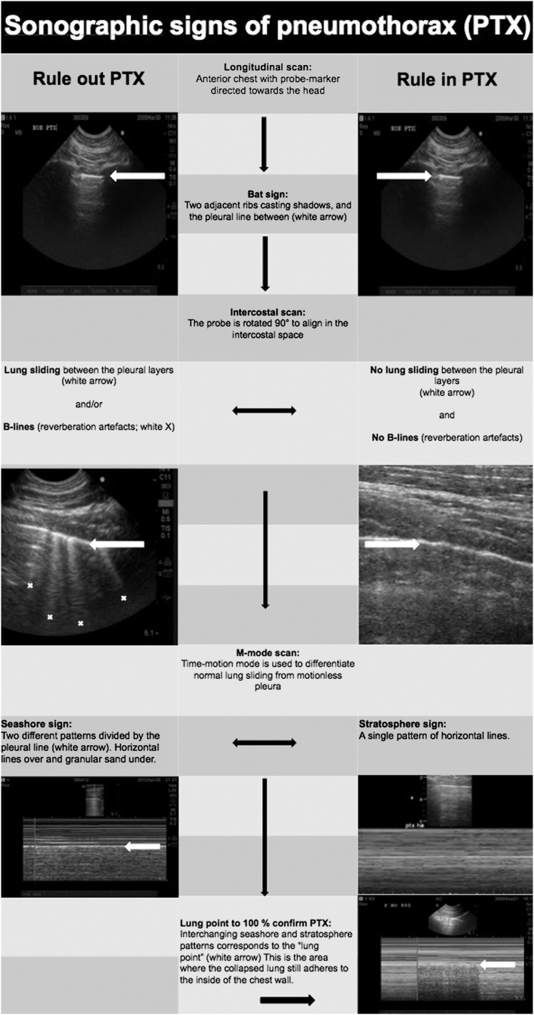
Figure 2.
Flowchart suggesting the correct sequence for ultrasound identification of the lung point. The lung point is localized at the interface between two distinct sonographic patterns that are synchronous with respiration: one with no lung sliding (the “stratosphere sign” in M-mode) and the other with normal lung sliding (the “seashore sign” in M-mode) illustrated in the lower right corner. Demonstration of normal lung sliding is only possible on video clips.

